The Microscope, in pictures
Six missions. Each one opens the next. Every picture below is the real microscope.
iWhat everything is
The eyepiece is on the left. Every control is in the panel on the right, and the knobs are under the eyepiece.
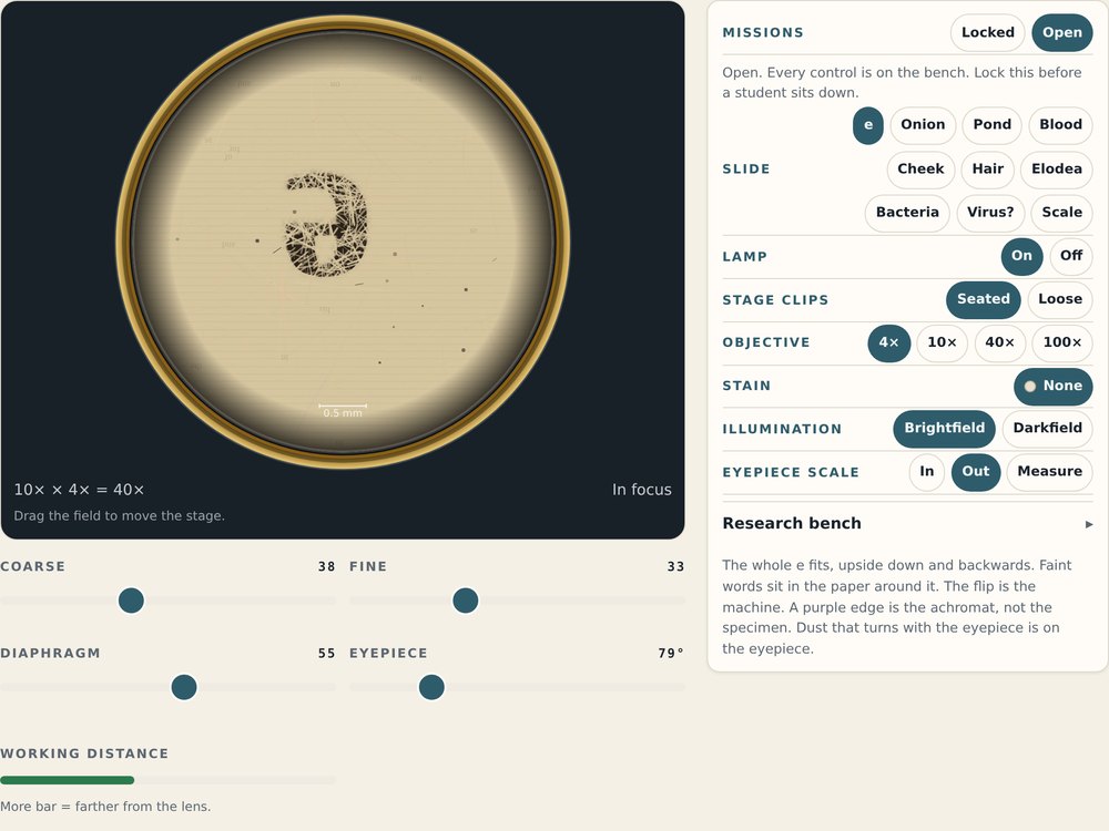
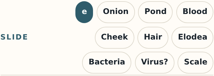 SlideWhat is under the lens. Start with the letter e.
 LampNo light, no picture.
 Stage clipsSeated holds the slide still.
 ObjectiveHow big. 4× is the smallest and the safest.
 CoarseBig focus moves. Only on 4× and 10×.
 FineSmall focus moves. The only knob on 40× and 100×.
 DiaphragmMore or less light. Fixes a washed-out picture.
 EyepieceTurns the eyepiece. Finds dust on the glass.
 Working distanceHow far the lens is from the glass. Red means too close.
 Missions (teacher)Locked opens one control at a time. Lock it before a student sits down.
1Make the microscope work Mission
Light, then the clips, then 4×, then the coarse knob.
Lamp: OnPress On.Clips: SeatedPress Seated.4×Start on the smallest.CoarseSlide it until the corner says In focus.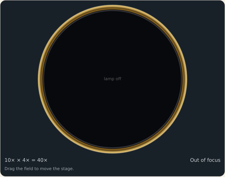 Lamp offBlack. Turn the lamp on.
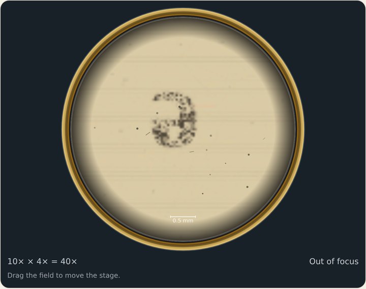 Out of focusKeep sliding Coarse.
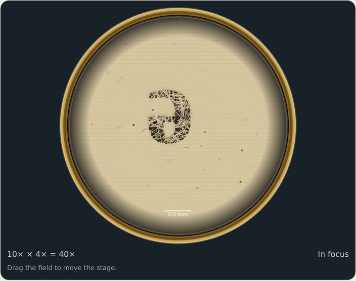 In focusDone.
The e is upside down and backwards. That is the lenses. It is not a mistake.
2See more Mission
10×, then 40×. On 40× use the fine knob only.
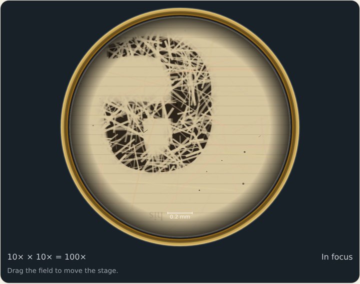 10×Bigger. Still use Coarse here.
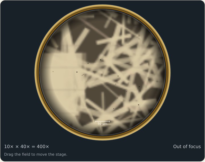 40×, blurryNow use Fine.
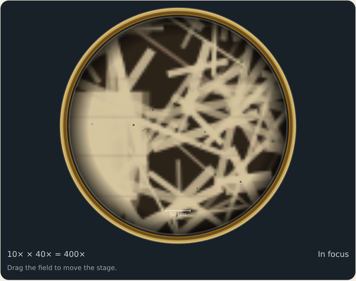 40×, sharpYou are inside the ink. Those are paper fibers.
DON'T
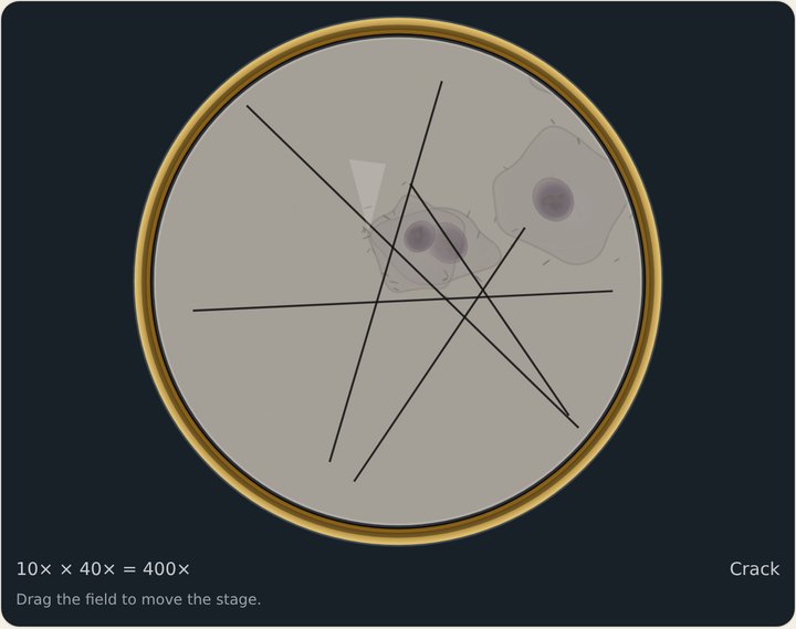 Coarse on 40× = crackThe lens hit the glass. Press New slide and start again on 4×.On 40× and 100×: Fine onlySmall moves. The picture is sharp only in a very thin zone.
3Change what you see Mission
Stain the onion. Then darkfield a live pond drop — with no stain.
Slide: OnionThen 40× and Fine. Stain: IodineThe stain buttons appear once a slide can take one.
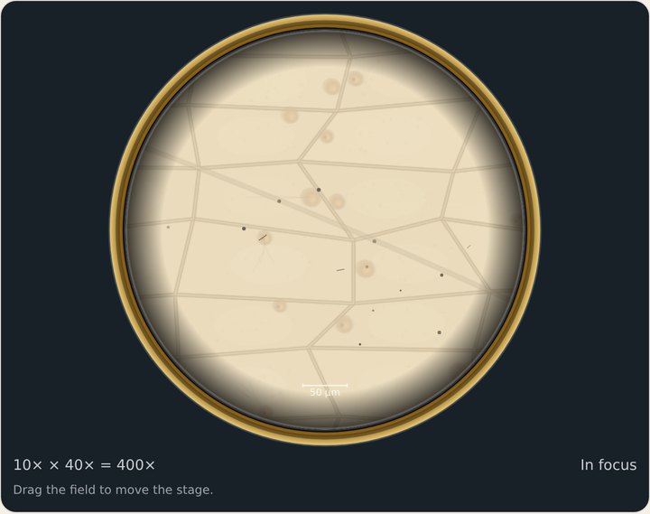 No stainA ghost. You can barely see the cells.
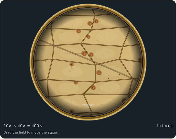 IodineThe walls go dark and every nucleus turns brown.
 Illumination: DarkfieldLoad Pond first. Stain: None.
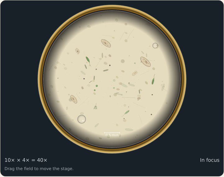 BrightfieldPale and hard to see.
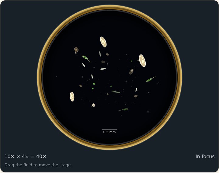 DarkfieldThe field goes black. Living things glow.
Why no stain on the pond? A stain kills what it colors. Darkfield lets you see them alive.
4Measure Mission
Scale in. Then Measure, and tap two points on a cell at 40×.
 Eyepiece scale: In, then MeasureThe ruler lives in the eyepiece, so it stays still when the slide moves.
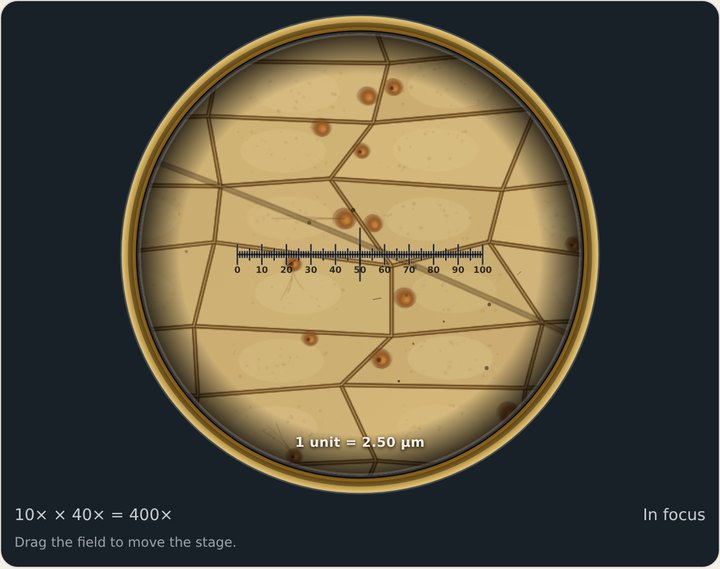 Scale inAt 40×, 1 unit = 2.5 µm.
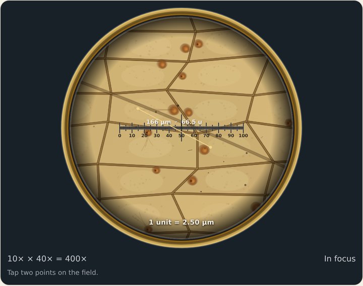 Two tapsTap one end of a cell, then the other. Read the number.
5What can’t this microscope see? Mission
Load Virus?, go to 100×, and put the oil on.
100×Fine knob only. Immersion oil: On the 100×This row appears on 100×.
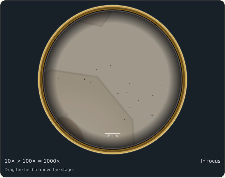 No oilDim and soft. That is physics, not a broken lens.
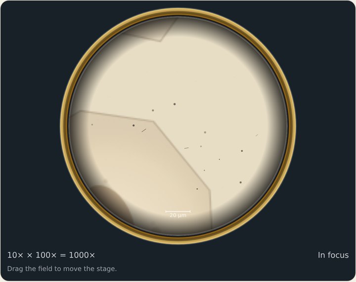 Oil onThe cells a virus lives in. No virus.
The honest ceiling. A virus is about 0.10 µm. Light cannot draw anything that small. 1,000× with oil is as far as this microscope goes.
6The research bench Mission
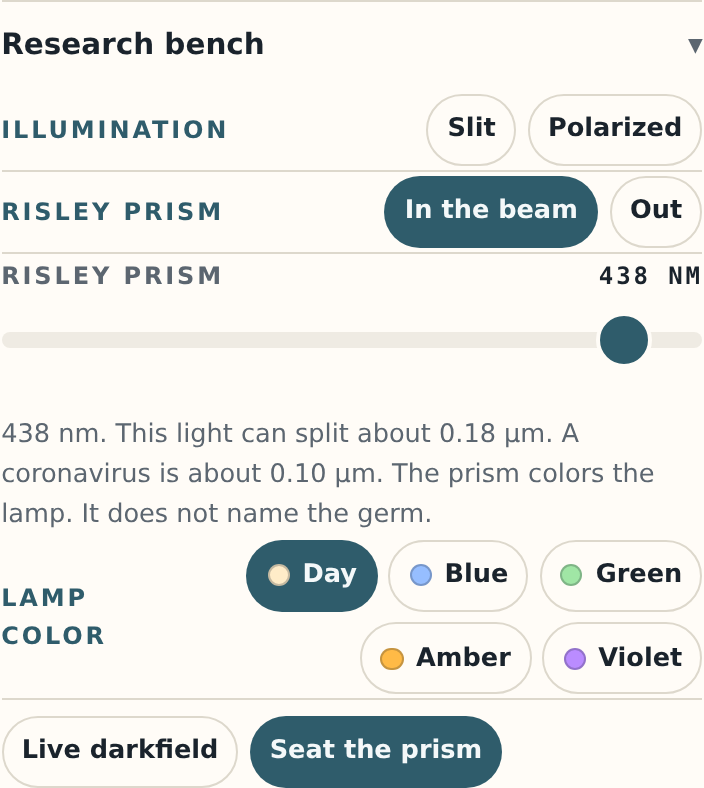
Open Research bench at the bottom of the panel. Slit, then polarized light, then the prism.
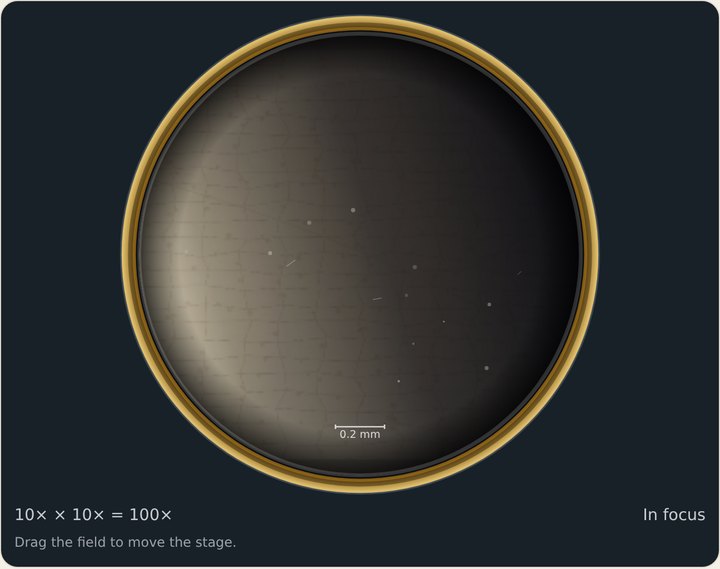 SlitLight from one side. Edges on the bright side catch it.
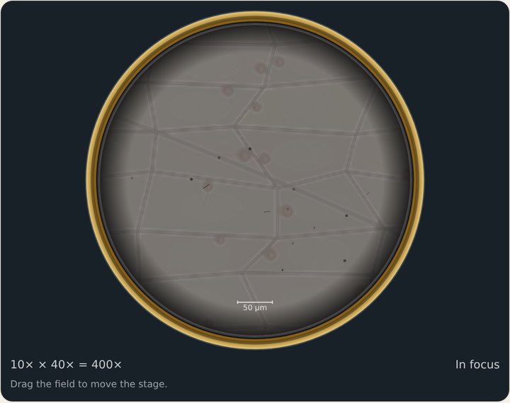 Polarized (onion)The field goes gray. The cell walls still show.
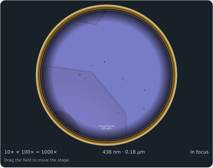 Prism on Virus?Read the number: about 0.18 µm. Still bigger than 0.10.
The prism changes the lamp’s color. It does not find the virus.
?Something looks wrong
- The circle is blackLamp: On. Then check the lamp before you blame the focus.
- It is blurry4× or 10×: Coarse. 40× or 100×: Fine only.
- Cracked glassCoarse on high power did it. Press New slide, start again on 4×.
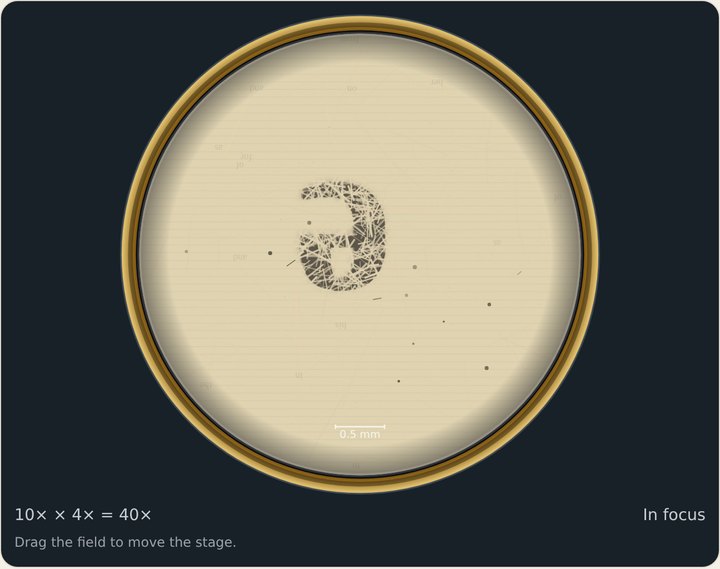 Too bright and washed outTurn the Diaphragm down.
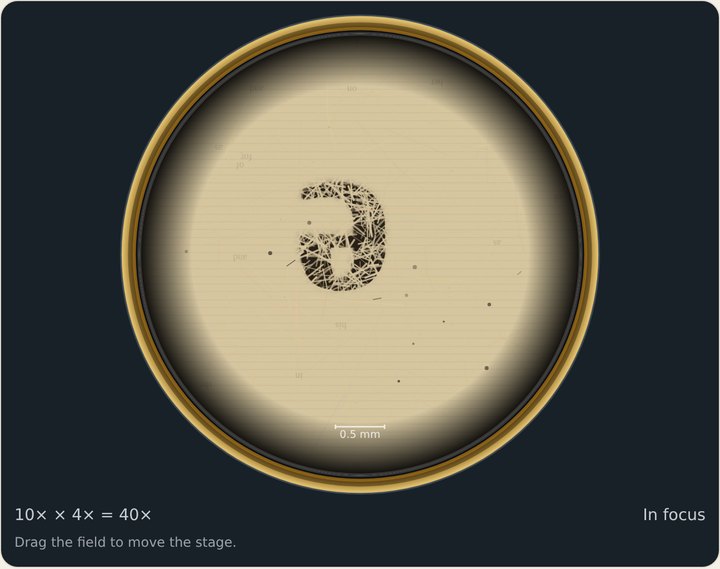 Too dark around the edgeTurn the Diaphragm up.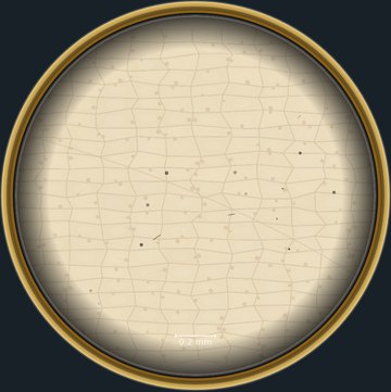
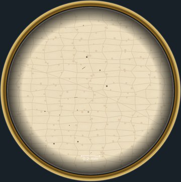 A speck that will not move when you dragTurn the Eyepiece knob. If the speck turns with it (compare the two pictures), it is dust on the eyepiece, not on the slide.
- Dim and soft on 100×Put the oil on.
- The slide keeps slidingStage clips: Seated.